Page History
...
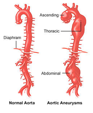
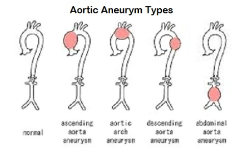
Types of Aortic Aneurysm
Renal Artery Aneurysm
There are many ways to ask if someone has an aneurysm. If the person has prior history of a particular type of aneurysm, say, aortic aneurysm, and the physician suspects that the patient suffers from another one, then specialized imaging instruments that are location-specific can be used. Examples are TRANSESOPHAGEAL ECHOCARDIOGRAPHY or TRANSTHORACIC ECHOCARDIOGRAPHY, they are used to look for abnormalities in the chest and the abdomen. The original data elements provided to us by DUKE and ACC, from which we created the examples in the CV-imaging TAUG, whether a patient has an aneurysm is asked in a pre-specified fashion. The questions would look like the following:
Does the patient have Aortic aneurysm? Aortic arch aneurysm? Thoracic aortic aneurysm? Suprarenal abdominal aneurysm? etc.
In this instance the specific anatomical location pre-specified in the question is where the test is performed.
...
| title | cv.xpt |
|---|---|
| Name | cv |
...
Row 3:
...
Indicates the presence of an aneurysm in the thoracic aorta.
...
Row 4:
...
Shows that the aneurysm led to the dissection of the thoracic aorta.
...
Row 5:
...
Shows the confirmed aortic dissection of the thoracic aorta from the previous assessment then undergoes anatomic classification. In this case, the patient's dissection is classified as Standford Class A, indicating that this aortic dissection involves the ascending aorta regardless of the site of origin [ref].
...
Rows 6-7:
...
Indicate the absence of aneurysms in both the suprarenal and infrarenal abdominal aorta.
...
Row
...
STUDYID
...
DOMAIN
...
USUBJID
...
CVGRPID
...
CVTESTCD
...
CVTEST
...
CVORRES
...
CVSTRESC
...
CVLOC
...
CVMETHOD
...
VISITNUM
...
VISIT
...
CVDTC
...
However, often times when patients complain about discomforts and pain, Often times when patients complain about chest discomforts and pain and the occurrence of aneurysm(s) is suspected, it is difficult to pinpoint the exact location of an the aneurysm hence . Hence MRI and CT scan are the most frequently used tools to detect aneurysmaneurysms. In this scenario, the test location is often broadly defined as the subject's chest, abdominal cavity or the whole-body. The specific location(s) where aneurysms are found are actually locational qualifiers for the results, not test.
| Dataset wrap | ||||||||||||||||||||||||||||||||||||||||||||||||||||||||||||||||||||||||||||||||||||||||||||||||||||||||||||||||||||||
|---|---|---|---|---|---|---|---|---|---|---|---|---|---|---|---|---|---|---|---|---|---|---|---|---|---|---|---|---|---|---|---|---|---|---|---|---|---|---|---|---|---|---|---|---|---|---|---|---|---|---|---|---|---|---|---|---|---|---|---|---|---|---|---|---|---|---|---|---|---|---|---|---|---|---|---|---|---|---|---|---|---|---|---|---|---|---|---|---|---|---|---|---|---|---|---|---|---|---|---|---|---|---|---|---|---|---|---|---|---|---|---|---|---|---|---|---|---|---|
| ||||||||||||||||||||||||||||||||||||||||||||||||||||||||||||||||||||||||||||||||||||||||||||||||||||||||||||||||||||||
|
...


