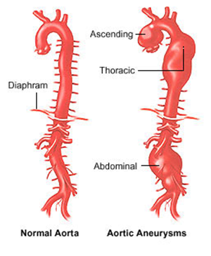
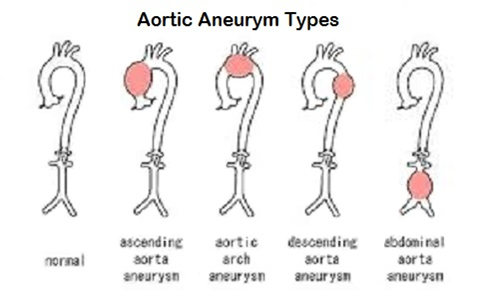
Types of Aortic Aneurysm


Renal Artery Aneurysm

Often times when patients complain about chest discomforts and pain and the occurrence of aneurysm(s) is suspected, it is difficult to pinpoint the exact location of the aneurysm. Hence MRI and CT scan are the most frequently used tools to detect aneurysms. In this scenario, the test location is often broadly defined as the subject's chest, abdominal cavity or the whole-body. The specific location(s) where aneurysms are found are actually locational qualifiers for the results, not test.
Rows 1-2: | Show subject ABC-123 has a single aortic aneurysm from a chest MRI scan. |
|---|
Row 3: | Shows the said aneurysm is 7.5cm in length (diameter), which is measured from the aortic arch to the abdominal aorta. |
|---|
| Rows 4-5: | Show subject ABC-456 is found to have aneurysms in two locations from a whole-body MRI Scan: RENAL ARTERY and THORACIC AORTA. |
|---|
|
Row | STUDYID | DOMAIN | USUBJID | CVSEQ | CVGRPID | CVTEST | CVORRES | CVORRESU | CVLOC | CVMETHOD | VISITNUM | VISIT | CVDTC |
| CVRESLOC1 | CVRESLOC2 | CVRLODTL |
|---|
| 1 | ABC | CV | ABC-123 | 1 | 1 | Aneurysm Indicator | Y |
| CHEST | MRI | 1 | BASELINE | 2020-04-27 |
|
|
|
|
|---|
| 2 | ABC | CV | ABC-123 | 2 | 1 | Number of Aneurysms | 1 |
| CHEST | MRI | 1 | BASELINE | 2020-04-27 |
| AORTA |
|
|
|---|
| 3 | ABC | CV | ABC-123 | 3 | 1 | Aneurysm Length/Diameter | 7.5 | CM | CHEST | MRI | 1 | BASELINE | 2020-04-27 |
| AORTA |
| Aortic Arch to Descending Aorta |
|---|
| 4 | ABC | CV | ABC-456 | 1 | 2 | Aneurysm Indicator | Y |
| BODY | MRI | 1 | BASELINE | 2020-04-27 |
|
|
|
|
|---|
| 5 | ABC | CV | ABC-456 | 2 | 2 | Number of Aneurysms | 2 |
| BODY | MRI | 1 | BASELINE | 2020-04-27 |
| RENAL ARTERY | THORACIC AORTA |
|
|---|
|
|
From the CV-imaging project we also encountered use-cases where we need sub-loc variables to help to further specify the detailed, more granular locations where a test is performed. Those values are indeed location information that should NOT be pre-coordinated into the TEST itself, but are also inappropriate for LOC.
So we need a variable that provides additional, more specific and granular information about a proximate location or a range of location(s).

This example shows the minor axis cross-sectional diameter measurements of the left and right ventricle of the heart, at end ventricular diastole.
Row 1: | Shows the cross-sectional diameter of the left ventricle at end ventricular diastole, measured along the minor axis and specifically at the location of the high papillary muscle. The further anatomical details at which the measurement is set and performed is represented by the CVLOCDTL NSV. |
|---|
Row 2: | Shows the cross-sectional diameter of the right ventricle at end ventricular diastole, measured along the minor axis and specifically at just below the tricuspid valve. The further anatomical details at which the measurement is set and performed is represented by the CVLOCDTL NSV. |
|---|
|
Row | STUDYID | DOMAIN | USUBJID | CVSEQ | CVTESTCD | CVTEST | CVORRES | CVORRESU | CVLOC | CVMETHOD | VISITNUM | VISIT | CVDTC |
| CVLOCDTL |
|---|
| 1 | ABC | CV | ABC-123 | 1 | MNDIAEVD | Minor Axis Cross-sec Diameter, EVD | 3.7 | CM | HEART, LEFT VENTRICLE | TTE | 1 | BASELINE |
|
| At high papillary muscle level |
|---|
| 2 | ABC | CV | ABC-123 | 2 | MNDIAEVD | Minor Axis Cross-sec Diameter, EVD | 3.2 | Cm | HEART, RIGHT VENTRICLE | TTE | 1 | BASELINE |
|
| Below the tricuspid valve |
|---|
|
|


